
Truncus Arteriosus: A Fetal Echocardiography Perspective
Dr. Mohammed Nizam Uddin, MBBS, BCS (Health), MD (Cardiology) Post Doctoral Training in Cardiac Intervention | Advanced Fellowship in Fetal Cardiology & Intervention Pioneer Fetal Cardiologist & Fetal Echocardiography Expert
Introduction
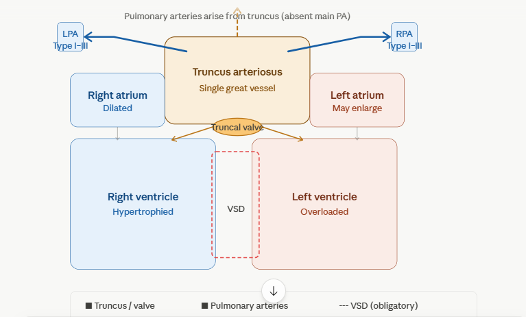
Truncus arteriosus (TA) is a conotruncal congenital heart defect characterized by a single arterial vessel arising from the base of the heart, giving origin to the aorta, pulmonary arteries, and coronary arteries through a single semilunar (truncal) valve. It accounts for approximately 1–3% of all congenital heart defects and carries significant mortality if undetected antenatally. Fetal echocardiography offers a critical window for early diagnosis, classification, and counselling before birth.
Embryological Basis
In normal cardiac development, the primitive truncus arteriosus undergoes septation between the 5th and 7th weeks of gestation, dividing into the aorta and main pulmonary artery. In truncus arteriosus, this aortopulmonary septation fails entirely, resulting in a single outflow vessel. This failure is commonly associated with abnormalities of neural crest cell migration and is strongly linked with chromosome 22q11.2 deletion (DiGeorge syndrome).
Classification (Van Praagh / Collett & Edwards)
The most widely used classification is based on the origin of the pulmonary arteries from the truncus:
Type I — A short main pulmonary artery segment arises from the truncus before dividing into right and left pulmonary arteries. This is the most common type (~50%).
Type II — The right and left pulmonary arteries arise separately but in close proximity from the posterior aspect of the truncus, with no main PA segment.
Type III — The right and left pulmonary arteries arise from lateral aspects of the truncus, more widely separated.
Type IV (Pseudo-truncus / Van Praagh Type A4) — Pulmonary blood supply comes from aortopulmonary collateral vessels; the main and branch PAs may be absent. This is now classified separately as pulmonary atresia with VSD.
Van Praagh A3 — One pulmonary artery is absent; one lung is supplied by a collateral.
Fetal Echocardiography: Systematic Approach
Scanning Protocol
A structured segmental approach is essential. The following views are used:
Four-chamber view — The four-chamber view may appear deceptively normal in early gestation. With advancing gestational age, abnormalities become more apparent including biventricular enlargement, right ventricular hypertrophy, and sometimes cardiomegaly.
Three-vessel and trachea (3VT) view — This is one of the most diagnostically sensitive views. The normal three-vessel arrangement (main PA, aorta, SVC) is replaced by a single large vessel (the truncus) and the SVC. The pulmonary artery is absent from its expected position. This is a near-pathognomonic finding and should trigger full cardiac evaluation.
Five-chamber and outflow tract views — A single large vessel is seen overriding the interventricular septum. The hallmark sign is the absence of two separate great vessels. Only one semilunar valve is identified — the truncal valve.
Short-axis view — Helps characterize truncal valve morphology, the number of leaflets, and the relationship of the coronary arteries.
Key Echocardiographic Features
1. Single Overriding Great Vessel
The most defining feature is a single arterial trunk arising from both ventricles, straddling a large perimembranous or outlet ventricular septal defect. On the long-axis view, the truncus appears significantly larger than either a normal aorta or pulmonary artery would individually be, reflecting the combined output of both ventricles.
2. Obligatory VSD
A large VSD is invariably present in all true cases of truncus arteriosus. It is almost always perimembranous or outlet in location, positioned directly beneath the truncal valve. Without this VSD, blood cannot exit the right ventricle. In fetal life, because both ventricles communicate through the VSD and contribute to truncal output, hemodynamic consequences are different from postnatal physiology.
3. Truncal Valve Abnormalities
The truncal valve replaces both the aortic and pulmonary valves. It may have 2, 3, 4, or even 5–6 leaflets. Tricuspid morphology is most common. Truncal valve regurgitation or stenosis can develop and must be carefully assessed using colour Doppler and spectral Doppler. Significant regurgitation may produce hydrops fetalis. Thickened or dysplastic leaflets suggest stenosis.
4. Origin of Pulmonary Arteries
Careful interrogation of the posterior and lateral walls of the truncus is essential to identify where the pulmonary arteries arise. In Type I, a short main PA segment is seen before bifurcation. In Types II and III, the branch PAs arise separately and may be challenging to trace. Colour flow mapping is invaluable in delineating PA origins.
5. Coronary Artery Origins
The coronary arteries arise from the truncus, typically near the truncal valve sinuses. Anomalous coronary origins — such as a single coronary, intramural course, or high take-off — are seen in a significant proportion of cases and carry important surgical implications.
6. Aortic Arch
In approximately 25–30% of cases, an interrupted aortic arch (IAA) coexists, most commonly Type B interruption between the left common carotid and left subclavian arteries. This combination is strongly associated with 22q11.2 deletion and must prompt genetic referral.
Doppler Findings in Fetal Echocardiography
Colour Doppler demonstrates turbulent, mixed flow within the single truncus. Both ventricular outflows converge into the truncus without separation. Regurgitant jets through the truncal valve, when present, are readily mapped.
Pulsed wave (PW) Doppler at the truncal valve shows a biphasic flow pattern — systolic forward flow with varying degrees of diastolic reversal in cases of significant regurgitation. Elevated peak velocity raises suspicion for truncal valve stenosis.
The branch pulmonary arteries show antegrade flow in fetal life, which is an important differentiating point from pulmonary atresia, where no antegrade flow is detectable within the pulmonary circulation.
Associated Anomalies
Truncus arteriosus is rarely an isolated lesion. Important associations include:
Chromosome 22q11.2 deletion — Present in 30–40% of cases, especially with conotruncal defects and interrupted aortic arch. Thymic hypoplasia, facial dysmorphism, and palatal anomalies may be detectable prenatally.
Interrupted aortic arch (Type B) — Significantly worsens postnatal prognosis and alters surgical planning.
Absent ductus arteriosus — Common in truncus arteriosus; the ductus is often absent or non-functional since the pulmonary arteries arise directly from the truncus.
Aberrant subclavian artery — Occasionally seen and may alter arch anatomy.
Extracardiac anomalies — Genitourinary, skeletal, and neurological malformations may coexist, particularly in chromosomal syndromes.
Differential Diagnosis on Fetal Echo
Tetralogy of Fallot with pulmonary atresia — A large, overriding aorta is seen, but the pulmonary valve remnant may be identifiable. The RV outflow is blind-ending. No separate truncal valve covers both outflows.
Double outlet right ventricle (DORV) — Both great arteries arise predominantly from the right ventricle, but two separate great vessels and semilunar valves are identifiable.
Transposition of the great arteries (TGA) — Two separate great vessels run in parallel; the aorta arises from the RV anteriorly and the PA from the LV posteriorly. Two valves are present.
Pulmonary atresia with VSD (pseudo-truncus) — The overriding aorta may mimic truncus, but no antegrade pulmonary artery flow is detectable. Pulmonary blood supply depends on aortopulmonary collaterals or a patent ductus.
Prognostic Considerations from Fetal Diagnosis
Antenatal diagnosis allows a multidisciplinary team — comprising obstetricians, fetal cardiologists, neonatologists, and cardiac surgeons — to formulate a birth plan. Delivery should be planned at a tertiary centre with pediatric cardiac surgery capability.
Fetal prognosis is influenced by the severity of truncal valve regurgitation, presence of IAA, coexistence of chromosomal anomalies, and associated extracardiac malformations. Significant truncal regurgitation may precipitate fetal cardiac failure, hydrops, and intrauterine death if severe.
Postnatal surgical correction — the Rastelli-type repair involving VSD closure directing LV output to the truncus, and insertion of a conduit from the RV to the pulmonary arteries — is performed in the neonatal period, ideally within the first 2–4 weeks of life. Early repair prevents irreversible pulmonary vascular disease from developing due to unrestricted pulmonary blood flow.
FCPS Cardiology Viva Pearls
The 3VT view is the single most important screening view for truncus arteriosus. Absence of the normal three-vessel arrangement with replacement by a single vessel should never be dismissed.
A single overriding vessel + absent separate pulmonary artery + large VSD = truncus arteriosus until proven otherwise.
Truncal valve regurgitation is the key hemodynamic determinant of fetal wellbeing. Serial fetal echos are mandatory once regurgitation is identified.
Always offer fetal karyotype / chromosomal microarray (CMA) given the strong association with 22q11.2 deletion.
Interrupted aortic arch must be actively excluded by tracing the complete aortic arch in every case of suspected truncus.
Absent ductus arteriosus is a pointer — its absence combined with a large single vessel should raise suspicion for truncus rather than pulmonary atresia.
Short Viva Answer
Q. What are the fetal echocardiographic features of truncus arteriosus?
Fetal echocardiography in truncus arteriosus reveals a single large great vessel (truncus) overriding a large VSD, arising from both ventricles. The 3VT view shows absence of the normal main pulmonary artery. A single truncal valve — which may be bicuspid, tricuspid, or quadricuspid — replaces both semilunar valves. Pulmonary arteries arise from the truncus, and Doppler confirms antegrade pulmonary flow. Associated features include truncal valve regurgitation, interrupted aortic arch in up to 30% of cases, and strong association with 22q11.2 deletion.
👨⚕️ Dr. Mohammed Nizam Uddin — MBBS, BCS (Health), MD (Cardiology) | Post Doctoral Training in Cardiac Intervention | Advanced Fellowship in Fetal Cardiology & Intervention | Associate Professor & Senior Consultant (Cardiology) | Pioneer Fetal Cardiologist & Fetal Echocardiography Expert